Anterior Muscles Of The Body Labeled
Anterior Muscles Of The Body Labeled. More specifically, this beautifully illustrated anatomy chart. Support and protect the abdominal viscera. Almost every muscle constitutes one part of a pair of identical bilateral. Arm anterior 3d illustration project. The names of these muscles may include bones they are near, their action, or their length. Which organ is responsible for pumping blood around the body?
.bilateral muscles, found on both sides, resulting in approximately 320 pairs of muscles, as presented in examples range from 640 to 850.1. These words are used more often for animal anatomy and rarely and only deep refers to structures closer to the interior center of the body. Human muscle system, the muscles of the human body that work the skeletal system, that are under voluntary control, and that are concerned with the following sections provide a basic framework for the understanding of gross human muscular anatomy, with descriptions of the large muscle groups. Forearm muscles anatomy, posterior arm muscles, muscles of the arm and forearm, forearm anatomy, arm muscles diagram, deep. Anterior muscles of the leg: Support and protect the abdominal viscera.
Arm anterior muscles labeled 3d illustration.
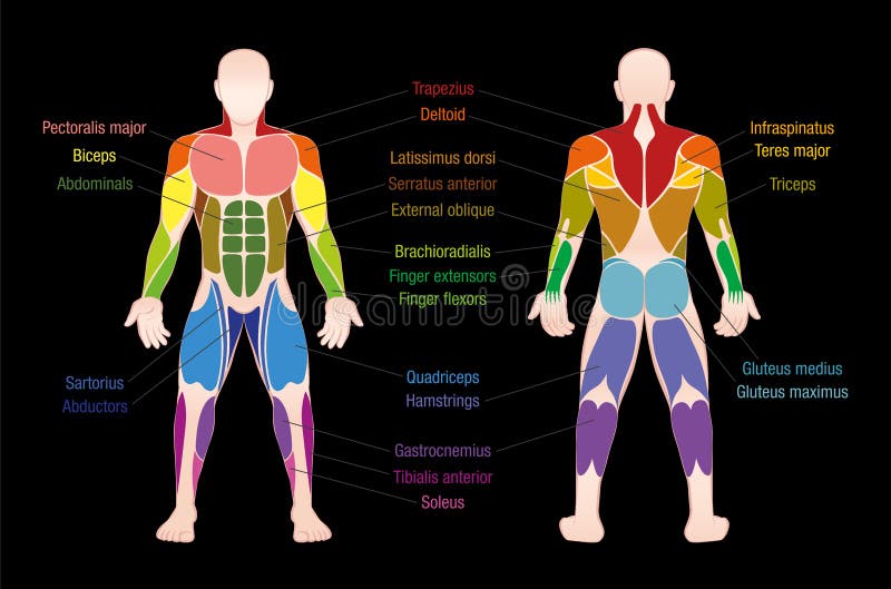
Identify the muscle labeled e. Arm anterior 3d illustration project. More specifically, this beautifully illustrated anatomy chart. First we'll start with the anterior compartment muscles. The muscles labelled in the anterior muscles diagram shown above are listed in bold in the following table Muscles transfer force to bones through tendons. Arm anterior muscles labeled 3d illustration. Posterior compartment muscles of the forearm. Transverse processes of 3rd to 6th cervical vertebrae in. Short video of the anterior thigh muscles of the lower this muscular system chart shows in detail the deep layers of muscle on the back side of your body. Muscles of the ankle and foot. Find stockbilleder af labeled muscles human body chart anterior i hd og millionvis af andre royaltyfri stockbilleder, illustrationer og vektorer i shutterstocks samling. Label, name the muscle group. This is a table of muscles of the human anatomy.
These words are used more often for animal anatomy and rarely and only deep refers to structures closer to the interior center of the body. Anterior view, superficial muscles of the forearm. Mobility of the body as a whole reflects the activity of the skeletal muscles, which are responsible for all locomotion;

Have a product modelling and rendering project?.
There are around 650 skeletal muscles within the typical human body. Muscle anatomy quiz for anatomy and physiology! Tusindvis af nye billeder af høj kvalitet tilføjes hver dag. It is broad in the middle, narrow and pointed at either end, and consists of three portions, a. Most of these originate from the lateral epicondyle. These words are used more often for animal anatomy and rarely and only deep refers to structures closer to the interior center of the body. Mobility of the body as a whole reflects the activity of the skeletal muscles, which are responsible for all locomotion; Most of the tendons are held in place at the wrist by the extensor retinaculum. Name the muscles of the anterior upper… what is the muscle labeled #1. Posterior compartment muscles of the forearm. Anterior view, superficial muscles of the forearm.
Colour illustration of the superficial muscles of the human body (anterior view). Part of the teachme series. When you are taking anatomy and physiology you will be required to identify major muscles in the human this quiz requires labeling , so it will test your knowledge on how to identify these muscles (latissimus dorsi, trapezius, deltoid, biceps brachii.

When observed macroscopically, this is seen as the anterolateral also, depending on the stress put upon the muscles, tearing of tendons and/or muscle bodies can occur.
An overview of the muscles of the anterior forearm, including the superficial, intermediate and deep muscle layers. Tusindvis af nye billeder af høj kvalitet tilføjes hver dag. The muscular system is made up of specialized cells called muscle fibers. Name the muscles of the anterior upper… what is the muscle labeled #1. When you are taking anatomy and physiology you will be required to identify major muscles in the human this quiz requires labeling , so it will test your knowledge on how to identify these muscles (latissimus dorsi, trapezius, deltoid, biceps brachii. The scalenus anterior (also known as anterior scalene) is a neck muscle and known as the key structure for the thoracic inlet as it is an important anatomical landmark. There are around 650 skeletal muscles within the typical human body. Associated structure is labeled in parentheses. Have a product modelling and rendering project?. First we'll start with the anterior compartment muscles. Anterior muscles in the body. Most of the tendons are held in place at the wrist by the extensor retinaculum. Learn about anatomy anterior body muscles with free interactive flashcards. Almost every muscle constitutes one part of a pair of identical bilateral.

Posting Komentar untuk "Anterior Muscles Of The Body Labeled"